The chapter about the anatomy of the Goodeidae is based on the contribution of Abraham Kobelkowsky to the book Viviparous Fishes from Mari Carmen Uribe and Harry Grier, 2005, New Life Publications (ISBN: 0-9645058-5-1), but structured a bit differently and supplemented by more details. The original title of this paper within the book is: "General Anatomy and Sexual Dimorphism of Goodea atripinnis (Teleostei: Goodeidae)", and is used as a basis for this article with the friendly permission of Mari Carmen Uribe, to who we communicate our thanks. This chapter deals exclusively with the species Goodea atripinnis, and consequently, not all things described herein can be assigned to other species, especially not to the Empetrichthyinae. Nevertheless, many things are universal valid for all species or genera, and therefore this chapter should serve as a brief introduction to the anatomy of this amazing group of fish.
Following mostly Abraham Kobelkowsky, we divide the chapter into the following subchapters:
1. The Skeleton
2. The Musculature
3. The Digestive System
4. The Urogenital System
5. The Circulatory System
6. The Nervous System and the Sense Organs
The Skeleton
The skeleton of Goodeid fishes (Goodeidae) can be divided into five bigger units:
1. The neurocranium sensu lato, bearing the brain and visual, olfactory and auditory organs.
2. An apparatus formed by the bones of the upper and the lower jaw, the bones of the mandibular suspension and the bones of the opercular series, all of them involved in feeding and breathing.
3. Paired fins, corresponding to our arms and legs, used mainly in manoeuvering.
4. The vertebral column or spine, including ribs and the caudal fin.
5. The unpaired dorsal and anal fin.
The neurocranium
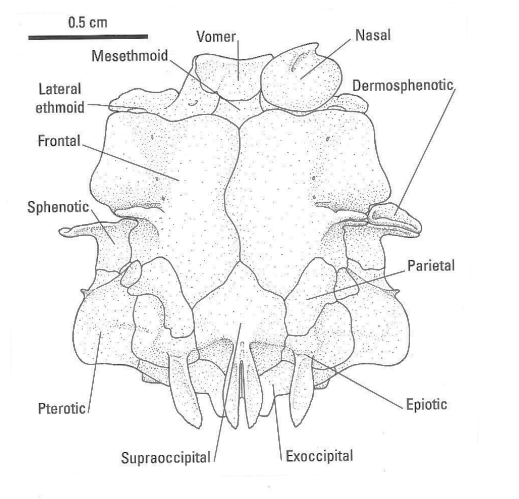
The neurocranium sensu lato is composed of two parts, meaning the neurocranium sensu stricto, which is composed of the bones providing direct support and protection to the brain and the visual, olfactory, and auditory organs, and bones that are intimately added to the neurocranium. These additional bones include the frontal, the parasphenoid, the parietal and the prevomer. In Goodea atripinnis, the neurocranium sensu lato is wide and dorsoventrally flattened.
Again, the major bones of the neurocranium sensu stricto occur in four groups:
1. The occipital region including the unpaired basioccipital and supraoccipital and the paired exoccipitals and epiotics.
2. The otic region (with prootic, intercalar and pterotic).
3. The suborbital region with only lacrimal and dermosphenotic.
4. The ethmoidal or olfactory region, including the mesethmoid and two lateral ethmoids.
The basioccipital is a median, unpaired bone forming the posterior base of the skull. It is the connection between skull and spine. Anteriorly, the unpaired parasphenoid in a discrete angle forms the roof of the mouth. The basioccipital is upwards attached to two exoccipitals, forming the posterior part of the skull. Dorsally and extending anteriorly, forming the roof of the braincase, the exoccipitals are connected to the unpaired supraoccipital and the paired exoccipitals or epiotics. The supraoccipital crest is divided into two posterior alary processes, receiving the muscles supracarinalis anteriores. Each epiotic develops a posterior fragile alary process receiving part of the musculature epiaxialis. The most prominent bones of the skull are the paired wide and asymmetrical frontals, extending from the supraoccipital anteriorly. Between frontals and epiotics are located two smaller parietals. The frontals are connected anteriorly with a pair of lateral ethmoids and the intermediate cartilaginous mesethmoid. With this olfactory region, the wide and laminar prevomer is connected, that is ventrally attached to the parasphenoid. Two nasals, oval and convex, covering the olfactory region.
From the suborbital series, only lacrimal (connected with the nasal) and dermosphenotic (joined to the frontal) are present, carrying part of the lateral line system. A pair of pterotics (posteriorly) and sphenotics (anteriorly) connect the dermosphenotic with epiotic and exoccipital. Each pterotic develops a wide shelf that includes the horizontal semicircular canal, and dorsally receives part of the axial musculature. Each sphenotic has a ventral process joining the dermosphenotic. Ventrally, a pair of prootics together with small intercalars complete together with the pterotics the otic region.


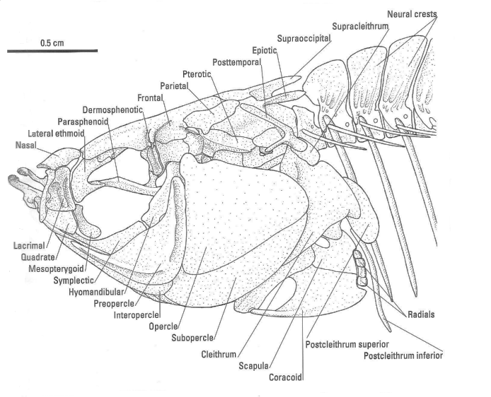
The drawing on the left side shows the dorsal view of the Neurocranium of Goodea atripinnis, the right one the left lateral view of the cephalic skeleton (A. Kobelkowsky).
The jaws, mandibular suspension, hyoid and branchial apparatus
The premaxillae, forming together with two maxillas the upper jaw, have a short ascending process and bear two kind of teeth. The dentary, which has an irregular shape is forming the most anterior part of the lower jaw, and also has got two types of teeth. They are arranged in two rows of marginal bifid teeth, and an ensemble of shorter, conic teeth. The other bones of the lower jaw are articular and angular. The articular has anteriorly an articular fossa, therefore forming the articulation to the quadrate, the first bone from the mandibular suspension.
The bones of the mandibular suspension are slender; the symplectic is remarkably large whereas the mesopterygoid is short, metapterygoid and ectopterygoid are absent. The hyomandibular, originally part of the second gill arch, connect the lower jaw with the skull.
Originated from the same second gill arch is the hyoid apparatus. It is composed of five branchiostegals and a group of unpaired, median bones. These branchiostegals support the branchiostegal or gill membrane, that encloses the gill chamber ventrally. The median bones of the hyoid arch are a small interhyal, a slender basihyal, a long and slender urohyal, a ceratohyal, connected to the first four branchistegals and an epihyal, connected to the fifth branchistegal.
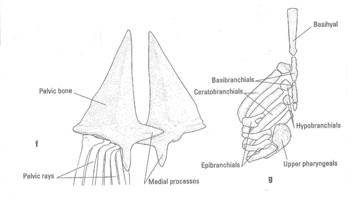
The branchial apparatus is formed by 3 slender basibranchials, 3 short hypobranchials, 4 long ceratobranchials, 4 epibranchials, the toothed lower pharyngeal and the 3 toothed pharyngobranchials. The last basibranchial is cartilaginous. The upper and lower pharyngeal teeth are pointed and vary gradually from robust to seriform.
The operculum is composed of two parts. Preopercle and interopercle cover the anterior region of the hyoid arch and parts of the mandibular suspension, opercle and subopercle cover the gill chamber with the branchial apparatus.
The spine and the fins
Functionally, the pectoral girdle may be seen as part of the head, supporting more or less the gill chamber posteriorly. The uppermost boneis a slender and forked posttemporal bone, that has a large superior process. The following supracleithrum is short, and from it originates Baudelots ligament and inserts on the basioccipital. The postcleithrum superior is oval and laminar, whereas the postcleithrum inferior is slender; both postcleithra contact the first pleural rib. Other important bones of the pectoral girdle are cleithrum, scapula and coracoid. Coracoid and scapula are connected by 4 radials (or actinosts) with the rays of the pectoral fin.
The pelvic girdle is represented by two single triangular bones, called pelvic bones, that have overlapping medial processes and carry 7 pelvic fin rays.
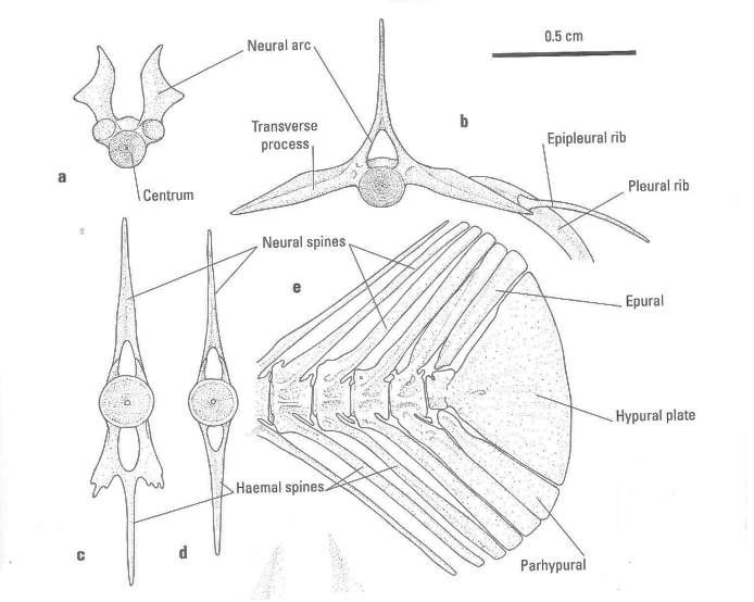
The spine or vertebral column can be divided into three groups of vertebrae. The anterior ones are called precaudal vertebrae, the posterior ones caudal vertebrae and the last part of the spine is formed by the caudal complex. The spine is connected to the basioccipital with the first of 19 or 20 precaudal vertebrae. This first precaudal vertebra has an open neural arch and lacks transverse processes. The remaining ones have wide transverse processes. Vertebrae 2 to 6 develop neural crests, whereas the rest bear neural spines. All except the first precaudal vertebra have got curved, strong pleural ribs on their transverse processes and 12 small and short epipleural ribs, articulating with the the first 11 pleural ribs. The first epipleural rib is corresponding to the first vertebra, but not contacting it.
The first caudal vertebra has one hemal arch with laminat, lateral processes. The others have spines instead of any processes.
The caudal complex includes one dorsally lying epural, the urostyle, the wide hypural plate and the ventrally lying parhypural. However, the neural and hemal processes of the last four vertebrae additionally support the caudal rays.
The dorsal fin is composed of 14 (13 in females) dorsal proximal pterygophores or radials, tiny distant radials and 14 (13 in females) dorsal rays; the anal fin by 15 anal proximal pterygophores or radials, tiny distant radials and 15 anal rays. The anal pterygophores are larger in males than in females. The anal fin pterygophore - unit is called andropodial (gonopodial) suspension. The largest male anal proximal pterygophore can be divided into the SL 8 times, but 10.8 times in females. In males, the first anal ray is very small, the following 5 rays are shortened and separated from the remaining 9 or 10 by a notch, forming a structure, called gonopodium or andropodium. In order to distinguish it from the Poeciliid gonopodium, that is marked differently in shape and function, we prefer the term andropodium for this part of the Goodeid reproductive organ.
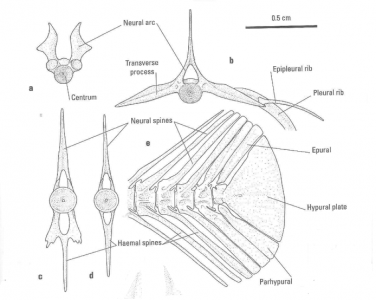

The drawings show the Skeleton of Goodea atripinnis: a) Anterior view of the first precaudal vertebra. b) Anterior view of the fifth vertebra, showing the articulation of ribs. c) First caudal vertebra. d) Fifth caudal vertebra. e) Caudal complex. f) Dorsal view of the pelvic girdle. g) Dorsal view of the branchial apparatus (A. Kobelkowsky).
The Musculature
The visceral musculature encompasses the muscles of the head and the gill archs. Among the visceral muscles of Goodea atripinnis, the largest muscle is the adductor mandibulae which is straight, oriented longitudinally and is subdivided into the bundles A1 and A2. It is responsible for closing the jaws. The most important muscle for opening the mouth is the sternohyoideus. Likewise, is an extensive muscle the adductor arcus palatini. It plays - together with the levator arcus palatini - an important role in expanding the oral cavity. The protractor hyoidei brings the lower jaw down.
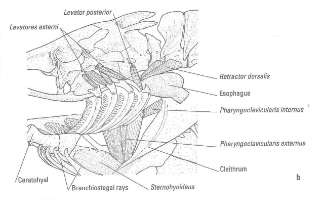
Based on their origin and insertion the dorsal branchial arch muscles can be divided into three groups: the external branchial arch levators, connecting the branchial arches to the neurocranium, called levatores externi I to IV for the first four branchial arches. The fifth arch has the levator posterior instead, well-separated from the levatores externi. The internal branchial arch levators or levatores interni are connecting the pharyngobranchials to the neurocranium. Finally the dorsal oblique muscles, or obliqui dorsales, are interconnecting the branchial arches and pharyngobranchials. Functionally, however, there are only two categories with the following properties: The first, which consists of the external branchial arch levators alone, is active during every respiratory cycle. These muscles expand the branchial basket through gill arch abduction and, in combination with hyomandibular pumping movements, lower the floor of the buccal cavity. The results of these combined movements are: The gill arches remain evenly distributed within the expanding branchial cavities during inspiration, so that continuity of the gill curtain is maintained. Water flow resistance is reduced also. The volume of water flowing into the buccal cavity during inspiration is increased. The second category, comprising the internal branchial arch levators and the dorsal oblique muscles, contracts only during the cough and else is completely inactive. Contraction of these muscles reinforces the dorsal suspension of the gill arches by firmly anchoring the pharyngobranchials and epibranchials to the base of the skull. In this way strong, caudally directed forces which develop during the intermediate expansion of the cough can be prevented from dislocating the branchial basket. An adductores series of five muscles connect the epibranchial and ceratobranchial elements of each arch.
Concerning the three muscles of the operculum, the dilatator operculi and levator operculi muscles are relatively broad. Together with the addcutor operculi, these muscles are responsible for opening and closing of the gill cover.
Pharyngeal musculature: A large muscle that extends posterodorsally from the upper pharyngeal jaw to the vertebral column is the retractor dorsalis muscle. It inserts on the two first precaudal vertebrae and is the dominant retractor of the upper pharyngeal jaw. An elongated muscle, the pharyngohyoideus serves primarily to protract the lower pharyngeal jaw. Acting as the functional antagonist of the pharyngohyoideus, the slender pharyngoclavicularis internus mainly mediates retraction and adduction of the lower pharyngeal jaw. The pharyngoclavicularis externus muscle has an especially wide insertion on the fifth ceratobranchial and functions mainly to mediate abduction of the lower pharyngeal jaw.
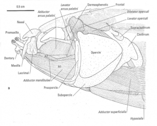

The drawings show the cephalic musculature of Goodea atripinnis: a) Left lateral view of the cheek musculature and pectoral fin. b) Left lateral view of the branchial musculature (A. Kobelkowsky).
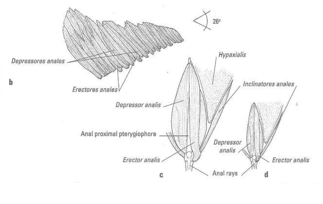
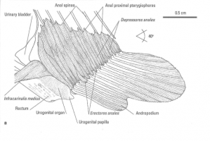
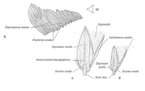
The musculature of the dorsal and anal fins consists of the inclinatores anales respectively dorsales, erectores anales respectively dorsales and depressores anales respectively dorsales. Whereas erectores and depressores are used to erect or depress the associated fin, the inclinatores move the fin rays sidewise and admit the fish to perform wavelike motions of the associated fins. The inclinatores anales muscles orginate from the fascia between the skin and the axial musculature; its longitude is bigger in males, occupying the 64.3% of the distance from the fin base to the horizontal septum, whereas in females it occupies 46.9%. The erectores anales originate from the lateral face of the proximal pterygiophores and insert widely on the anterolateral part of base of the rays; those of males are wider and longer. These muscles are broader near their insertions. The upper part of the first erector is more massive than the rest of erectors and the muscle inserts by a tendon on the first anal rays. The depressores anales muscles originate from the proximal pterygiophores and insert on the posterior part of the anal rays bases; those of males are larger.
Anterior to the anal musculature, and upon the urogenital organ, the infracarinalis medius muscle originates and surrounds part of the spermatic and urinary ducts of males. In females this muscle surrounds the urinary duct and oviduct. It is thicker in males.
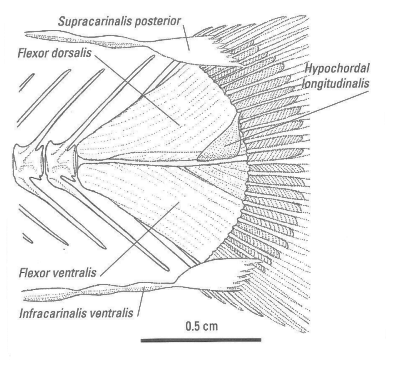
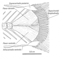
From the caudal musculature, the muscles flexor ventralis and flexor dorsalis are wide. They originate from the last 4 centra and hemal and neural spines, respectively. The flexor ventralis muscle inserts on the lateral bases of the ventral branched rays, whereas the flexor dorsalis inserts on the dorsal rays. The hypochordal longitudinalis muscle originates on the hypural plate and inserts on dorsal rays 4 and 5. Contractions of these 3 muscles lead to an outfanning of the caudal fin. The supracarinalis posterior and infracarinalis posterior muscles run posteriorly in the shape of small fans on the dorsal and ventral procurrent rays. They help in performing wavelike movements with the caudal fin.
The epiaxiales form the dorsalmost components of the body musculature, whereas the hypaxiales form the ventral mass of the body musculature and are situated below the horizontal septum. Coupled with the epiaxiales, the caudal components of the hypaxiales play a central role in propulsive locomotion.
The Digestive System
Goodea atripinnis has a horizontal mouth that is dorsally orientated. The premaxillary and dentary marginal teeth are arranged in two rows of setiform and bifid elements, followed of a nonossified matrix. A set of small conical teeth is on the roof and the floor of the bucal cavity. The bucopharyngal cavity shows the oral valve and lacks teeth on the palate, tongue and gill rakers. The gill rakers average 47 and are laminar and triangular.
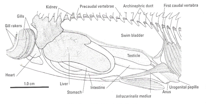
The esophagus is especially wide, whereas the stomach is slender and long. There are no pyloric caeca. The coiling pattern of the intestine is concentric along the right side of the liver, posterior to which it is straight. The length of the digestive tract is more than 230% of the standard length.
On the left side, the liver has a short and a large lobe, bordering the gall bladder. Dorsally the spleen is between the stomach and the intestinal loops.
The swim bladder is unicameral and extends from the renal lobes until the posterior part of the visceral cavity; it shows the rete mirabile in the anterior ventral part.

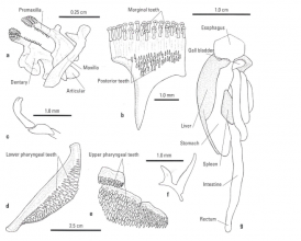
The left drawing shows the celomic cavities of Goodea atripinnis, left lateral view, and the right one
The Urogenital System
The kidneys are fused, composed of two wide lobes surrounding the esophagus. Both renal lobes are dorsally separated by a notch, through which the retractores dorsalis pass from the pharyngobranchials to the two first vertebrae. Each lobe contacts the sinus venosus of the heart.
The rest of the kidneys extends along the precaudal vertebrae. Since the renal mass occupies the middle part of each vertebral centrum, the ventral aspect of the kidney is stair-like.
The two archinephric ducts start from the posterior face of each renal lobe and run separately along the ventral part of precaudal vertebrae, the anterior part of the first anal spine, and the anal fin musculature. Both ducts join the urinary bladder, which has a funnel shape and continues posteroventrally as the common urinary duct inside the urogenital organ. This opens outside by the urinary opening in the female; in the male the common urinary duct fuses with the common spermatic duct to become the urogenital duct inside the urogenital organ, which exits via the urogenital opening. The urogenital papilla of G. atripinnis is bilobed, being shorter in females and longer and straight in males.
Ovaries fuse together with a tendency to have the left larger, and the right wider. In G. atripinnis the longitudinal septum separating the ovaries is complete. The oviduct or female gonoduct is relatively short and exits via the genital opening, immediately preceding the urinary opening.
The testes are elongate and fused together along the anterior third, which is rugged and pigmented. Inside each testsi, the main testicular duct is located near the medial faces and exits posteriorly as spermatic duct.
The Circulary System
The heart has a relatively short sinus venosus, which is hidden by the renal lobes; the auricle is assymetrical and the ventricle is triangular. The ventral aorta is diagonal and branches anteriorly in two pairs of afferent branchial anteries, and posteriorly into two other pairs. Dorsal to the branchial arches, the efferent branchial arteries 1 and 2 join together, as well as arteries 3 and 4, forming the two roots that fuse to give rise to the dorsal aorta.
The dorsal aorta is located along the left side of the vertebral centra. The celiac artery arises from the right side of the beginning of the dorsal aorta, marking the kidney with a ventral groove. This artery courses dorsally along the esophagus and continues until the digestive tract.
From the caudal vein, two veins arise and descend parallel to the archinephric ducts. The yrun anteriorly but lateroventrally to the swim bladder until the reach the renal lobes.
The Nervous System and the Sense Organs
The pores of the lateral line are concentrated on the dorsolateral part of the head. The olfactory organ is flat and shows just a discrete folding. The eye has the six ocular muscles, innervated by the corresponding cranial nerves. The optic nerve enters the cerebral hemispheres anteroventrally. The inner ear has a small utriculus; the sacculus contains the large sagitta and the lagena the small asteriscus.
The brain has anteriorly the olfactory bulbs, from which the olfactory nerves diverge until reaching the olfactory organs. The cerebral hemispheres are of medium size. The pineal organ is long and slender, and is oriented anteriorly. The optic lobes are conspicuous. The cerebellum forms laterally the auricular lobes. The medula oblongata dorsally has the tela choroidea and is more slender to form the medulla.
1. The neurocranium sensu lato, bearing the brain and visual, olfactory and auditory organs,
2. An apparatus formed by the bones of the upper and the lower jaw, the bones of the mandibular suspension and the bones of the opercular series, all of them involved in feeding
and breathing.
3. Paired fins, corresponding to our arms and legs, used mainly in manoeuvering.
4. The vertebral column or spine, including ribs and the caudal fin.
5. The unpaired dorsal and anal fin.
The neurocranium
The neurocranium sensu lato is composed of two parts, meaning the neurocranium sensu stricto, which is composed of the bones providing direct support and protection to the brain and the visual, olfactory, and auditory organs, and bones that are intimately added to the neurocranium. These additional bones include the frontal, the parasphenoid, the parietal and the prevomer. In Goodea atripinnis, the neurocranium sensu lato is wide and dorsoventrally flattened.
Again, the major bones of the neurocranium sensu stricto occur in four groups:
1. The occipital region including the unpaired basioccipital and supraoccipital and the paired exoccipitals and epiotics.
2. The otic region (with prootic, intercalar and pterotic).
3. The suborbital region with only lacrimal and dermosphenotic.
4. The ethmoidal or olfactory region, including the mesethmoid and two lateral ethmoids.
The basioccipital is a median, unpaired bone forming the posterior base of the skull. It is the connection between skull and spine. Anteriorly, the unpaired parasphenoid in a discrete angle forms the roof of the mouth. The basioccipital is upwards attached to two exoccipitals, forming the posterior part of the skull. Dorsally and extending anteriorly, forming the roof of the braincase, the exoccipitals are connected to the unpaired supraoccipital and the paired exoccipitals or epiotics. The supraoccipital crest is divided into two posterior alary processes, receiving the muscles supracarinalis anteriores. Each epiotic develops a posterior fragile alary process receiving part of the musculature epiaxialis. The most prominent bones of the skull are the paired wide and asymmetrical frontals, extending from the supraoccipital anteriorly. Between frontals and epiotics are located two smaller parietals. The frontals are connected anteriorly with a pair of lateral ethmoids and the intermediate cartilaginous mesethmoid. With this olfactory region, the wide and laminar prevomer is connected, that is ventrally attached to the parasphenoid. Two nasals, oval and convex, covering the olfactory region.
From the suborbital series, only lacrimal (connected with the nasal) and dermosphenotic (joined to the frontal) are present, carrying part of the lateral line system. A pair of pterotics (posteriorly) and sphenotics (anteriorly) connect the dermosphenotic with epiotic and exoccipital. Each pterotic develops a wide shelf that includes the horizontal semicircular canal, and dorsally receives part of the axial musculature. Each sphenotic has a ventral process joining the dermosphenotic. Ventrally, a pair of prootics together with small intercalars complete together with the pterotics the otic region.
The jaws, mandibular suspension, hyoid and branchial apparatus
The premaxillae, forming together with two maxillas the upper jaw, have a short ascending process and bear two kind of teeth. The dentary, which has an irregular shape is forming the most anterior part of the lower jaw, and also has got two types of teeth. They are arranged in two rows of marginal bifid teeth, and an ensemble of shorter, conic teeth. The other bones of the lower jaw are articular and angular. The articular has anteriorly an articular fossa, therefore forming the articulation to the quadrate, the first bone from the mandibular suspension.
The bones of the mandibular suspension are slender; the symplectic is remarkably large whereas the mesopterygoid is short, metapterygoid and ectopterygoid are absent. The hyomandibular, originally part of the second gill arch, connect the lower jaw with the skull.
Originated from the same second gill arch is the hyoid apparatus. It is composed of five branchiostegals and a group of unpaired, median bones. These branchiostegals support the branchiostegal or gill membrane, that encloses the gill chamber ventrally. The median bones of the hyoid arch are a small interhyal, a slender basihyal, a long and slender urohyal, a ceratohyal, connected to the first four branchistegals and an epihyal, connected to the fifth branchistegal.
The branchial apparatus is formed by 3 slender basibranchials, 3 short hypobranchials, 4 long ceratobranchials, 4 epibranchials, the toothed lower pharyngeal and the 3 toothed pharyngobranchials. The last basibranchial is cartilaginous. The upper and lower pharyngeal teeth are pointed and vary gradually from robust to seriform.
The operculum is composed of two parts. Preopercle and interopercle cover the anterior region of the hyoid arch and parts of the mandibular suspension, opercle and subopercle cover the gill chamber with the branchial apparatus.
The spine and the fins
Functionally, the pectoral girdle may be seen as part of the head, supporting more or less the gill chamber posteriorly. The uppermost boneis a slender and forked posttemporal bone, that has a large superior process. The following supracleithrum is short, and from it originates Baudelots ligament and inserts on the basioccipital. The postcleithrum superior is oval and laminar, whereas the postcleithrum inferior is slender; both postcleithra contact the first pleural rib. Other important bones of the pectoral girdle are cleithrum, scapula and coracoid. Coracoid and scapula are connected by 4 radials (or actinosts) with the rays of the pectoral fin.
The pelvic girdle is represented by two single triangular bones, called pelvic bones, that have overlapping medial processes and carry 7 pelvic fin rays.
The spine or vertebral column can be divided into three groups of vertebrae. The anterior ones are called precaudal vertebrae, the posterior ones caudal vertebrae and the last part of the spine is formed by the caudal complex. The spine is connected to the basioccipital with the first of 19 or 20 precaudal vertebrae. This first precaudal vertebra has an open neural arch and lacks transverse processes. The remaining ones have wide transverse processes. Vertebrae 2 to 6 develop neural crests, whereas the rest bear neural spines. All except the first precaudal vertebra have got curved, strong pleural ribs on their transverse processes and 12 small and short epipleural ribs, articulating with the the first 11 pleural ribs. The first epipleural rib is corresponding to the first vertebra, but not contacting it.
The first caudal vertebra has one hemal arch with laminat, lateral processes. The others have spines instead of any processes.
The caudal complex includes one dorsally lying epural, the urostyle, the wide hypural plate and the ventrally lying parhypural. However, the neural and hemal processes of the last four vertebrae additionally support the caudal rays.
The dorsal fin is composed of 14 dorsal proximal pterygophores or radials, tiny distant radials and 14 dorsal rays; the anal fin by 15 anal proximal pterygophores or radials, tiny distant radials and 15 anal rays. The anal pterygophores are larger in males than in females. The anal fin pterygophore - unit is called andropodial (gonopodial) suspension. The largest male anal proximal pterygophore can be divided into the SL 8 times, but 10.8 times in females. In males, the first anal ray is very small, the following 5 rays are shortened and separated from the remaining 9 or 10 by a notch, forming a structure, called gonopodium or andropodium. In order to distinguish it from the Poeciliid gonopodium, that is marked different in shape and function, we prefer the term andropodium for this part of the Goodeid reproductive organ.
skeleton of Goodeid fishes (Goodeidae) can be divided into five bigger units:
1. The neurocranium sensu lato, bearing the brain and visual, olfactory and auditory organs,
2. An apparatus formed by the bones of the upper and the lower jaw, the bones of the mandibular suspension and the bones of the opercular series, all of them involved in feeding
and breathing.
3. Paired fins, corresponding to our arms and legs, used mainly in manoeuvering.
4. The vertebral column or spine, including ribs and the caudal fin.
5. The unpaired dorsal and anal fin.
The neurocranium
The neurocranium sensu lato is composed of two parts, meaning the neurocranium sensu stricto, which is composed of the bones providing direct support and protection to the brain and the visual, olfactory, and auditory organs, and bones that are intimately added to the neurocranium. These additional bones include the frontal, the parasphenoid, the parietal and the prevomer. In Goodea atripinnis, the neurocranium sensu lato is wide and dorsoventrally flattened.
Again, the major bones of the neurocranium sensu stricto occur in four groups:
1. The occipital region including the unpaired basioccipital and supraoccipital and the paired exoccipitals and epiotics.
2. The otic region (with prootic, intercalar and pterotic).
3. The suborbital region with only lacrimal and dermosphenotic.
4. The ethmoidal or olfactory region, including the mesethmoid and two lateral ethmoids.
The basioccipital is a median, unpaired bone forming the posterior base of the skull. It is the connection between skull and spine. Anteriorly, the unpaired parasphenoid in a discrete angle forms the roof of the mouth. The basioccipital is upwards attached to two exoccipitals, forming the posterior part of the skull. Dorsally and extending anteriorly, forming the roof of the braincase, the exoccipitals are connected to the unpaired supraoccipital and the paired exoccipitals or epiotics. The supraoccipital crest is divided into two posterior alary processes, receiving the muscles supracarinalis anteriores. Each epiotic develops a posterior fragile alary process receiving part of the musculature epiaxialis. The most prominent bones of the skull are the paired wide and asymmetrical frontals, extending from the supraoccipital anteriorly. Between frontals and epiotics are located two smaller parietals. The frontals are connected anteriorly with a pair of lateral ethmoids and the intermediate cartilaginous mesethmoid. With this olfactory region, the wide and laminar prevomer is connected, that is ventrally attached to the parasphenoid. Two nasals, oval and convex, covering the olfactory region.
From the suborbital series, only lacrimal (connected with the nasal) and dermosphenotic (joined to the frontal) are present, carrying part of the lateral line system. A pair of pterotics (posteriorly) and sphenotics (anteriorly) connect the dermosphenotic with epiotic and exoccipital. Each pterotic develops a wide shelf that includes the horizontal semicircular canal, and dorsally receives part of the axial musculature. Each sphenotic has a ventral process joining the dermosphenotic. Ventrally, a pair of prootics together with small intercalars complete together with the pterotics the otic region.
The jaws, mandibular suspension, hyoid and branchial apparatus
The premaxillae, forming together with two maxillas the upper jaw, have a short ascending process and bear two kind of teeth. The dentary, which has an irregular shape is forming the most anterior part of the lower jaw, and also has got two types of teeth. They are arranged in two rows of marginal bifid teeth, and an ensemble of shorter, conic teeth. The other bones of the lower jaw are articular and angular. The articular has anteriorly an articular fossa, therefore forming the articulation to the quadrate, the first bone from the mandibular suspension.
The bones of the mandibular suspension are slender; the symplectic is remarkably large whereas the mesopterygoid is short, metapterygoid and ectopterygoid are absent. The hyomandibular, originally part of the second gill arch, connect the lower jaw with the skull.
Originated from the same second gill arch is the hyoid apparatus. It is composed of five branchiostegals and a group of unpaired, median bones. These branchiostegals support the branchiostegal or gill membrane, that encloses the gill chamber ventrally. The median bones of the hyoid arch are a small interhyal, a slender basihyal, a long and slender urohyal, a ceratohyal, connected to the first four branchistegals and an epihyal, connected to the fifth branchistegal.
The branchial apparatus is formed by 3 slender basibranchials, 3 short hypobranchials, 4 long ceratobranchials, 4 epibranchials, the toothed lower pharyngeal and the 3 toothed pharyngobranchials. The last basibranchial is cartilaginous. The upper and lower pharyngeal teeth are pointed and vary gradually from robust to seriform.
The operculum is composed of two parts. Preopercle and interopercle cover the anterior region of the hyoid arch and parts of the mandibular suspension, opercle and subopercle cover the gill chamber with the branchial apparatus.
The spine and the fins
Functionally, the pectoral girdle may be seen as part of the head, supporting more or less the gill chamber posteriorly. The uppermost boneis a slender and forked posttemporal bone, that has a large superior process. The following supracleithrum is short, and from it originates Baudelots ligament and inserts on the basioccipital. The postcleithrum superior is oval and laminar, whereas the postcleithrum inferior is slender; both postcleithra contact the first pleural rib. Other important bones of the pectoral girdle are cleithrum, scapula and coracoid. Coracoid and scapula are connected by 4 radials (or actinosts) with the rays of the pectoral fin.
The pelvic girdle is represented by two single triangular bones, called pelvic bones, that have overlapping medial processes and carry 7 pelvic fin rays.
The spine or vertebral column can be divided into three groups of vertebrae. The anterior ones are called precaudal vertebrae, the posterior ones caudal vertebrae and the last part of the spine is formed by the caudal complex. The spine is connected to the basioccipital with the first of 19 or 20 precaudal vertebrae. This first precaudal vertebra has an open neural arch and lacks transverse processes. The remaining ones have wide transverse processes. Vertebrae 2 to 6 develop neural crests, whereas the rest bear neural spines. All except the first precaudal vertebra have got curved, strong pleural ribs on their transverse processes and 12 small and short epipleural ribs, articulating with the the first 11 pleural ribs. The first epipleural rib is corresponding to the first vertebra, but not contacting it.
The first caudal vertebra has one hemal arch with laminat, lateral processes. The others have spines instead of any processes.
The caudal complex includes one dorsally lying epural, the urostyle, the wide hypural plate and the ventrally lying parhypural. However, the neural and hemal processes of the last four vertebrae additionally support the caudal rays.
The dorsal fin is composed of 14 dorsal proximal pterygophores or radials, tiny distant radials and 14 dorsal rays; the anal fin by 15 anal proximal pterygophores or radials, tiny distant radials and 15 anal rays. The anal pterygophores are larger in males than in females. The anal fin pterygophore - unit is called andropodial (gonopodial) suspension. The largest male anal proximal pterygophore can be divided into the SL 8 times, but 10.8 times in females. In males, the first anal ray is very small, the following 5 rays are shortened and separated from the remaining 9 or 10 by a notch, forming a structure, called gonopodium or andropodium. In order to distinguish it from the Poeciliid gonopodium, that is marked different in shape and function, we prefer the term andropodium for this part of the Goodeid reproductive organThe skeleton of Goodeid fishes (Goodeidae) can be divided into five bigger units:
1. The neurocranium sensu lato, bearing the brain and visual, olfactory and auditory organs,
2. An apparatus formed by the bones of the upper and the lower jaw, the bones of the mandibular suspension and the bones of the opercular series, all of them involved in feeding
and breathing.
3. Paired fins, corresponding to our arms and legs, used mainly in manoeuvering.
4. The vertebral column or spine, including ribs and the caudal fin.
5. The unpaired dorsal and anal fin.
The neurocranium
The neurocranium sensu lato is composed of two parts, meaning the neurocranium sensu stricto, which is composed of the bones providing direct support and protection to the brain and the visual, olfactory, and auditory organs, and bones that are intimately added to the neurocranium. These additional bones include the frontal, the parasphenoid, the parietal and the prevomer. In Goodea atripinnis, the neurocranium sensu lato is wide and dorsoventrally flattened.
Again, the major bones of the neurocranium sensu stricto occur in four groups:
1. The occipital region including the unpaired basioccipital and supraoccipital and the paired exoccipitals and epiotics.
2. The otic region (with prootic, intercalar and pterotic).
3. The suborbital region with only lacrimal and dermosphenotic.
4. The ethmoidal or olfactory region, including the mesethmoid and two lateral ethmoids.
The basioccipital is a median, unpaired bone forming the posterior base of the skull. It is the connection between skull and spine. Anteriorly, the unpaired parasphenoid in a discrete angle forms the roof of the mouth. The basioccipital is upwards attached to two exoccipitals, forming the posterior part of the skull. Dorsally and extending anteriorly, forming the roof of the braincase, the exoccipitals are connected to the unpaired supraoccipital and the paired exoccipitals or epiotics. The supraoccipital crest is divided into two posterior alary processes, receiving the muscles supracarinalis anteriores. Each epiotic develops a posterior fragile alary process receiving part of the musculature epiaxialis. The most prominent bones of the skull are the paired wide and asymmetrical frontals, extending from the supraoccipital anteriorly. Between frontals and epiotics are located two smaller parietals. The frontals are connected anteriorly with a pair of lateral ethmoids and the intermediate cartilaginous mesethmoid. With this olfactory region, the wide and laminar prevomer is connected, that is ventrally attached to the parasphenoid. Two nasals, oval and convex, covering the olfactory region.
From the suborbital series, only lacrimal (connected with the nasal) and dermosphenotic (joined to the frontal) are present, carrying part of the lateral line system. A pair of pterotics (posteriorly) and sphenotics (anteriorly) connect the dermosphenotic with epiotic and exoccipital. Each pterotic develops a wide shelf that includes the horizontal semicircular canal, and dorsally receives part of the axial musculature. Each sphenotic has a ventral process joining the dermosphenotic. Ventrally, a pair of prootics together with small intercalars complete together with the pterotics the otic region.
The jaws, mandibular suspension, hyoid and branchial apparatus
The premaxillae, forming together with two maxillas the upper jaw, have a short ascending process and bear two kind of teeth. The dentary, which has an irregular shape is forming the most anterior part of the lower jaw, and also has got two types of teeth. They are arranged in two rows of marginal bifid teeth, and an ensemble of shorter, conic teeth. The other bones of the lower jaw are articular and angular. The articular has anteriorly an articular fossa, therefore forming the articulation to the quadrate, the first bone from the mandibular suspension.
The bones of the mandibular suspension are slender; the symplectic is remarkably large whereas the mesopterygoid is short, metapterygoid and ectopterygoid are absent. The hyomandibular, originally part of the second gill arch, connect the lower jaw with the skull.
Originated from the same second gill arch is the hyoid apparatus. It is composed of five branchiostegals and a group of unpaired, median bones. These branchiostegals support the branchiostegal or gill membrane, that encloses the gill chamber ventrally. The median bones of the hyoid arch are a small interhyal, a slender basihyal, a long and slender urohyal, a ceratohyal, connected to the first four branchistegals and an epihyal, connected to the fifth branchistegal.
The branchial apparatus is formed by 3 slender basibranchials, 3 short hypobranchials, 4 long ceratobranchials, 4 epibranchials, the toothed lower pharyngeal and the 3 toothed pharyngobranchials. The last basibranchial is cartilaginous. The upper and lower pharyngeal teeth are pointed and vary gradually from robust to seriform.
The operculum is composed of two parts. Preopercle and interopercle cover the anterior region of the hyoid arch and parts of the mandibular suspension, opercle and subopercle cover the gill chamber with the branchial apparatus.
The spine and the fins
Functionally, the pectoral girdle may be seen as part of the head, supporting more or less the gill chamber posteriorly. The uppermost boneis a slender and forked posttemporal bone, that has a large superior process. The following supracleithrum is short, and from it originates Baudelots ligament and inserts on the basioccipital. The postcleithrum superior is oval and laminar, whereas the postcleithrum inferior is slender; both postcleithra contact the first pleural rib. Other important bones of the pectoral girdle are cleithrum, scapula and coracoid. Coracoid and scapula are connected by 4 radials (or actinosts) with the rays of the pectoral fin.
The pelvic girdle is represented by two single triangular bones, called pelvic bones, that have overlapping medial processes and carry 7 pelvic fin rays.
The spine or vertebral column can be divided into three groups of vertebrae. The anterior ones are called precaudal vertebrae, the posterior ones caudal vertebrae and the last part of the spine is formed by the caudal complex. The spine is connected to the basioccipital with the first of 19 or 20 precaudal vertebrae. This first precaudal vertebra has an open neural arch and lacks transverse processes. The remaining ones have wide transverse processes. Vertebrae 2 to 6 develop neural crests, whereas the rest bear neural spines. All except the first precaudal vertebra have got curved, strong pleural ribs on their transverse processes and 12 small and short epipleural ribs, articulating with the the first 11 pleural ribs. The first epipleural rib is corresponding to the first vertebra, but not contacting it.
The first caudal vertebra has one hemal arch with laminat, lateral processes. The others have spines instead of any processes.
The caudal complex includes one dorsally lying epural, the urostyle, the wide hypural plate and the ventrally lying parhypural. However, the neural and hemal processes of the last four vertebrae additionally support the caudal rays.
The dorsal fin is composed of 14 dorsal proximal pterygophores or radials, tiny distant radials and 14 dorsal rays; the anal fin by 15 anal proximal pterygophores or radials, tiny distant radials and 15 anal rays. The anal pterygophores are larger in males than in females. The anal fin pterygophore - unit is called andropodial (gonopodial) suspension. The largest male anal proximal pterygophore can be divided into the SL 8 times, but 10.8 times in females. In males, the first anal ray is very small, the following 5 rays are shortened and separated from the remaining 9 or 10 by a notch, forming a structure, called gonopodium or andropodium. In order to distinguish it from the Poeciliid gonopodium, that is marked different in shape and function, we prefer the term andropodium for this part of the Goodeid reproductive organ.The skeleton of Goodeid fishes (Goodeidae) can be divided into five bigger units:
1. The neurocranium sensu lato, bearing the brain and visual, olfactory and auditory organs,
2. An apparatus formed by the bones of the upper and the lower jaw, the bones of the mandibular suspension and the bones of the opercular series, all of them involved in feeding
and breathing.
3. Paired fins, corresponding to our arms and legs, used mainly in manoeuvering.
4. The vertebral column or spine, including ribs and the caudal fin.
5. The unpaired dorsal and anal fin.
The neurocranium
The neurocranium sensu lato is composed of two parts, meaning the neurocranium sensu stricto, which is composed of the bones providing direct support and protection to the brain and the visual, olfactory, and auditory organs, and bones that are intimately added to the neurocranium. These additional bones include the frontal, the parasphenoid, the parietal and the prevomer. In Goodea atripinnis, the neurocranium sensu lato is wide and dorsoventrally flattened.
Again, the major bones of the neurocranium sensu stricto occur in four groups:
1. The occipital region including the unpaired basioccipital and supraoccipital and the paired exoccipitals and epiotics.
2. The otic region (with prootic, intercalar and pterotic).
3. The suborbital region with only lacrimal and dermosphenotic.
4. The ethmoidal or olfactory region, including the mesethmoid and two lateral ethmoids.
The basioccipital is a median, unpaired bone forming the posterior base of the skull. It is the connection between skull and spine. Anteriorly, the unpaired parasphenoid in a discrete angle forms the roof of the mouth. The basioccipital is upwards attached to two exoccipitals, forming the posterior part of the skull. Dorsally and extending anteriorly, forming the roof of the braincase, the exoccipitals are connected to the unpaired supraoccipital and the paired exoccipitals or epiotics. The supraoccipital crest is divided into two posterior alary processes, receiving the muscles supracarinalis anteriores. Each epiotic develops a posterior fragile alary process receiving part of the musculature epiaxialis. The most prominent bones of the skull are the paired wide and asymmetrical frontals, extending from the supraoccipital anteriorly. Between frontals and epiotics are located two smaller parietals. The frontals are connected anteriorly with a pair of lateral ethmoids and the intermediate cartilaginous mesethmoid. With this olfactory region, the wide and laminar prevomer is connected, that is ventrally attached to the parasphenoid. Two nasals, oval and convex, covering the olfactory region.
From the suborbital series, only lacrimal (connected with the nasal) and dermosphenotic (joined to the frontal) are present, carrying part of the lateral line system. A pair of pterotics (posteriorly) and sphenotics (anteriorly) connect the dermosphenotic with epiotic and exoccipital. Each pterotic develops a wide shelf that includes the horizontal semicircular canal, and dorsally receives part of the axial musculature. Each sphenotic has a ventral process joining the dermosphenotic. Ventrally, a pair of prootics together with small intercalars complete together with the pterotics the otic region.
The jaws, mandibular suspension, hyoid and branchial apparatus
The premaxillae, forming together with two maxillas the upper jaw, have a short ascending process and bear two kind of teeth. The dentary, which has an irregular shape is forming the most anterior part of the lower jaw, and also has got two types of teeth. They are arranged in two rows of marginal bifid teeth, and an ensemble of shorter, conic teeth. The other bones of the lower jaw are articular and angular. The articular has anteriorly an articular fossa, therefore forming the articulation to the quadrate, the first bone from the mandibular suspension.
The bones of the mandibular suspension are slender; the symplectic is remarkably large whereas the mesopterygoid is short, metapterygoid and ectopterygoid are absent. The hyomandibular, originally part of the second gill arch, connect the lower jaw with the skull.
Originated from the same second gill arch is the hyoid apparatus. It is composed of five branchiostegals and a group of unpaired, median bones. These branchiostegals support the branchiostegal or gill membrane, that encloses the gill chamber ventrally. The median bones of the hyoid arch are a small interhyal, a slender basihyal, a long and slender urohyal, a ceratohyal, connected to the first four branchistegals and an epihyal, connected to the fifth branchistegal.
The branchial apparatus is formed by 3 slender basibranchials, 3 short hypobranchials, 4 long ceratobranchials, 4 epibranchials, the toothed lower pharyngeal and the 3 toothed pharyngobranchials. The last basibranchial is cartilaginous. The upper and lower pharyngeal teeth are pointed and vary gradually from robust to seriform.
The operculum is composed of two parts. Preopercle and interopercle cover the anterior region of the hyoid arch and parts of the mandibular suspension, opercle and subopercle cover the gill chamber with the branchial apparatus.
The spine and the fins
Functionally, the pectoral girdle may be seen as part of the head, supporting more or less the gill chamber posteriorly. The uppermost boneis a slender and forked posttemporal bone, that has a large superior process. The following supracleithrum is short, and from it originates Baudelots ligament and inserts on the basioccipital. The postcleithrum superior is oval and laminar, whereas the postcleithrum inferior is slender; both postcleithra contact the first pleural rib. Other important bones of the pectoral girdle are cleithrum, scapula and coracoid. Coracoid and scapula are connected by 4 radials (or actinosts) with the rays of the pectoral fin.
The pelvic girdle is represented by two single triangular bones, called pelvic bones, that have overlapping medial processes and carry 7 pelvic fin rays.
The spine or vertebral column can be divided into three groups of vertebrae. The anterior ones are called precaudal vertebrae, the posterior ones caudal vertebrae and the last part of the spine is formed by the caudal complex. The spine is connected to the basioccipital with the first of 19 or 20 precaudal vertebrae. This first precaudal vertebra has an open neural arch and lacks transverse processes. The remaining ones have wide transverse processes. Vertebrae 2 to 6 develop neural crests, whereas the rest bear neural spines. All except the first precaudal vertebra have got curved, strong pleural ribs on their transverse processes and 12 small and short epipleural ribs, articulating with the the first 11 pleural ribs. The first epipleural rib is corresponding to the first vertebra, but not contacting it.
The first caudal vertebra has one hemal arch with laminat, lateral processes. The others have spines instead of any processes.
The caudal complex includes one dorsally lying epural, the urostyle, the wide hypural plate and the ventrally lying parhypural. However, the neural and hemal processes of the last four vertebrae additionally support the caudal rays.
The dorsal fin is composed of 14 dorsal proximal pterygophores or radials, tiny distant radials and 14 dorsal rays; the anal fin by 15 anal proximal pterygophores or radials, tiny distant radials and 15 anal rays. The anal pterygophores are larger in males than in females. The anal fin pterygophore - unit is called andropodial (gonopodial) suspension. The largest male anal proximal pterygophore can be divided into the SL 8 times, but 10.8 times in females. In males, the first anal ray is very small, the following 5 rays are shortened and separated from the remaining 9 or 10 by a notch, forming a structure, called gonopodium or andropodium. In order to distinguish it from the Poeciliid gonopodium, that is marked different in shape and function, we prefer the term andropodium for this part of the Goodeid reproductive organ.